Dry Eye Disease in Clinical
Practice

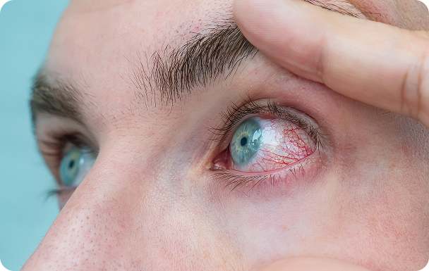
Dry eye syndrome is a multifactorial pathology of the ocular surface characterised by the loss of tear film homeostasis, and is accompanied by ocular symptoms in which tear film instability, hyperosmolarity, inflammation and induced damage of the ocular surface and neurosensory abnormalities play an important aetiological role1,2,3

The TFOS DEWS II Epidemiology Report (widely cited in dry eye research) summarizes evidence showing **worldwide prevalence estimates range from about 5% to 50%, depending on geographic region and diagnostic criteria.

Dry eye disease affects hundreds of millions of people around the world and is one of the main causes of visits to the ophthalmologist1

RISK FACTORS

REFRACTIVE SURGERY
PROLONGED USE OF ELETRONIC DEVICES
REDUCED INTAKE OF OMEGA 3
ENVIRONMENTAL FACTORS
PROLONGED USE OF CONTACT LENSES
FEMALES
SMOKING
AGEING
SYSTEMIC DRUGS
ENVIRONMENTAL FACTORS
REFRACTIVE SURGERY
PROLONGED USE OF ELETRONIC DEVICES
REDUCED INTAKE OF OMEGA 3
ENVIRONMENTAL FACTORS
PROLONGED USE OF CONTACT LENSES
FEMALES
SMOKING
AGEING
SYSTEMIC DRUGS
ENVIRONMENTAL FACTORS

Causes

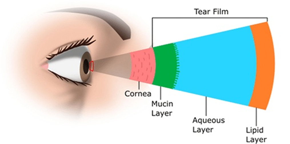
Dry eye disease can be caused by a variety of factors that disrupt the healthy tear film, which consists of

three layers, from inside to outside, Mucin, Aqueous and Lipid.

Three layers, from inside to outside, Mucin, Aqueous and Lipid.

Common causes include:

HYPOLACRIMATION

Quantitative defect in aqueous component of tears

DYSFUNCTIONAL LACRIMATION

Quantitative or qualitative defect of mucous or lipid component (like dysfunction of the meibomian glands that leads to excessive evaporation).

NORMAL TEARS

iso-osmolar tears

Normal concentration of solutes

TEARS IN A PATIENT WITH DRY EYES

Hyperosmolar tears

High concentration of solutes and decrease in volume

SymptomS

DED may cause a wide range of ocular symptoms including:

This is typically accompanied by fluctuating vision with blurred or double vision. These symptoms can significantly impact patients’ quality of life and lead to reduced work and learning efficiency.

DIAGNOSIS

Assessment of the symptoms:

OSDI questionnaire: assessment of the symptoms and impact on QoL, 12 questions with a score of 0-4 (never-always)
Results – score obtained x25 and :12:

0-12

13-22

23-32

33-100

Normal eye

Mild dry eye

Medium dry eye

Severe dry eye

Assessment of signs:

Ocular surface staining

Using eye stains like fluorescein or lissamine green for evaluation of ocular surface integrity and tear film stability

Schirmer's test

Assesses the tear volume by placing a small strip of paper under the lower eyelid

TBUT test

Measures tear film stability

Treatment

Managing dry eye disease often requires a combination of treatments tailored to the underlying cause.

Common treatments include:

PHASE I:

PHASE II:

PHASE III:

Dry eye disease is a multifactorial condition that can significantly impact quality of life1. Early diagnosis and appropriate management are crucial for preventing complications and maintaining eye health2. If you experience symptoms of dry eye, consult an eye care professional for a comprehensive evaluation and personalized treatment plan.

Advancing Eye Care Through a Complete Product Portfolio

Evidence-based solutions designed to support clinical excellence

References:
1. Stapleton, F., Alves, M., Bunya, V. Y., Jalbert, I., Lekhanont, K., Malet, F., Na, K.-S., Schaumberg, D., Uchino, M., Vehof, J., Viso, E., Vitale, S., & Jones, L. (2017). TFOS DEWS II Epidemiology Report. The Ocular Surface, 15(3), 334–365.

2. Craig, J. P., Nichols, K. K., Akpek, E. K., Caffery, B., Dua, H. S., Joo, C.-K., Liu, Z., Nelson, J. D., Nichols, J. J., Tsubota, K., & Stapleton, F. (2017). TFOS DEWS II Definition and Classification Report. The Ocular Surface, 15(3), 276–283.
3. Bron, A. J., de Paiva, C. S., Chauhan, S. K., Bonini, S., Gabison, E. E., Jain, S., Knop, E., Markoulli, M., Ogawa, Y., Perez, V. L., Sanchis-Gimeno, J. A., Schaumberg, D. A., Uchino, M., Vehof, J., Willcox, M. D., & Stapleton, F. (2017). TFOS DEWS II Pathophysiology Report. The Ocular Surface, 15(3), 438–510.


You are about to leave this website

External content may be subject to different local regulations, product availability may vary by market.

THIS PAGE IS INTENDED ONLY FOR HEALTHCARE PROFESSIONALS

To access this page, please click YES if you are a healthcare professional.
If you are not a healthcare professional, select NO.